
ADVANCED MAGNETIC TECHNOLOGIES AND CONSULTING (AMT&C)
Russia
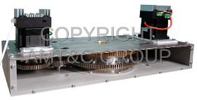
The magnetic system of this field source is also built on the principle of nested cylindrical Halbach-structures. An important feature is the presence of two motors, controlled independently, so it is possible to manage the change not only the magnitude but also the direction of the magnetic field. Because the device was designed for operation together with an optical microscope, the vertical size of the system near the working region is reduced to 90 mm. Stepper motors, that turn the two magnetic subsystems, are located on the sides of the casing
OSTEC ARTTOOL LIMITED COMPANY
Russia
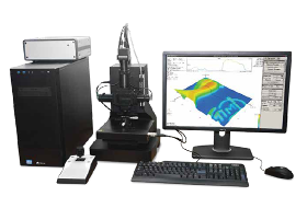
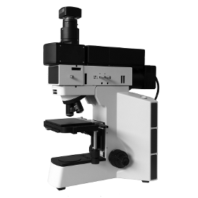
IMOS Interference Microscope-nanoprofilometer enables accurate, quantitative, ISOcompliant noncontact surface measurements and characterization of micro and nanoscale surface characteristics, capturing up to two million data points in seconds. Choosing the right optical profilometer depends on your application requirements, including speed, accuracy, vertical range, automation and flexibility. IMOS optical surface profilometer provides powerful versatility in noncontact optical surface measurements. The system makes it easy and fast to investigate a wide range of surface types, including smooth, rough, flat, sloped and stepped surfaces. All measurements are nondestructive, fast and require no special sample preparation.
NT-MDT LLC
Russia
IR s‑SNOM microscopy and spectroscopy with 10 nm spatial resolution Wide spectral range of operation 312 μm Incredibly low thermal drift and high signal stability Versatile AFM with advanced modes SRI (conductivity), KPFM (surface potential), SCM (capacitance), MFM (magnetic properties), PFM (piezoelectric forces) HybriD Mode™ quantitative nanomechanical mapping Integration with microRaman (optional) The ability of s‑SNOM measurements in the visible spectral range (optional) NTMDT Spectrum Instruments presents NTEGRA Nano IR scattering scanning nearfield optical microscope (s‑SNOM) designed for infrared (IR) spectral range. AFM probe is located in the focus of optical system which excites sample structure by IR laser and collects the optical response. Collected light is directed to Michelson interferometer for optical analysis.
NT-MDT LLC
Russia
Industry leading automation level Outstanding noise floor and thermal drifts Fast scanner with XYZ lownoise closeloop Routine atomic resolution 60+ SPM modes in basic configuration Continuous zoom from millimeter to nanometer range Integrated with new Atomic Force Microscopy technique HybriD Mode™ Atomic Force Microscope NEXT provides motorized sample positioning and integrated high resolution optical microscope positioning, motorized continuous zoom and focusing of the optical microscope. But AFM automation is more than just motorization. Powerful Nova PX software algorithms remove a gap between optics and AFM providing continuous zoom from huge panoramic optical view down to atomic resolution. Since all step movers are coupled together with the optical image, NEXT provides autofocus, fast oneclick cantilever alignment, panoramic optical view and multiple scanning on 5×5 mm range.Cantilever recognition and automatic laser alignment both in liquid and air Autofocus
TEKNIK CIZGI KESIM BUKUM METAL SAN VE TIC LTD STI
Turkey

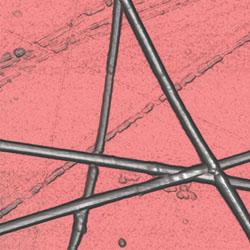
Materials - Metallography laboratory includes metallographic sample preparation equipment, 500X optical microscope, grain structure imaging and analysis program, hardness measuring devices.

Do you sell or make similar products?
Sign up to europages and have your products listed
LEICA MICROSYSTEMS
Germany
Visually inspect and chemically analyze in a single work step with your DM6 M LIBS materials analysis solution. The integrated laser spectroscopy function delivers the chemical composition of the microstructure that you see in the microscope image – within a second. Speed up your workflow. The LIBS module turns a Leica optical microscope into a 1-step solution that combines visual inspection and chemical analysis right at your workspace. Determine the composition of what you have visually identified within seconds. Use LIBS to perform advanced material analysis 90% faster compared to inspection with SEM/EDS. Surface contamination or coatings can also be easily removed. Chemical mapping and micro-drilling are further analytical steps.
OSTEC ARTTOOL LIMITED COMPANY
Russia
RAMOS S120 compact dual-channel confocal Raman microscope is designed for micro spectral measurements with capabilities at the level of high-end systems. RAMOS S120 microscope has a rigid, moving parts free design that requires no adjustments, has both high sensitivity and high spatial resolution, and can be equipped with two single-mode lasers simultaneously, 488/633 nm or 532/785 nm. Wide possibilities, high reliability, and compactness allow using RAMOS S120 for solving a wide range of scientific and industrial applications. Main features: - Research level optical microscope with advanced measurement techniques - Submicron resolution due to confocal design - One or two integrated single-mode lasers - Fully automated change of lasers/gratings without additional system alignment - Automatic adjustment of laser power - Wide dynamic range and extremely high sensitivity of innovative sCMOS detector - Fiber optic Raman probe option - Raman mapping with motorized sample stage
OSTEC ARTTOOL LIMITED COMPANY
Russia
RAMOS E/M series Raman spectrometers are designed on the basis of research-grade optical microscopes allowing the realization of the following light microscopy methods: - Raman measurements - Transmitted light - Reflected light (bright field and dark field illumination) - Confocal microscopy - Fluorescence measurements - Polarization contrast and phase-contrast imaging - Differential interference contrast 3D scanning laser Raman microscopes provide rapid, high sensitivity analysis. The innovative approach to system design of Raman spectrometers ensures extremely high temperature and temporal stability of spectral measurements. All RAMOS E200 system components are fully integrated within an optical microscope providing compactness and mobility of the system. In RAMOS M350, M520, M750 systems external imaging spectrographs are connected via optical fibers. Raman measurements with the RAMOS E/M Series systems can be started in several minutes by turning a system key.
OSTEC ARTTOOL LIMITED COMPANY
Russia
RAMOS U120 compact single-channel confocal Raman microscope is designed for micro spectral measurements with capabilities at the level of high-end systems. RAMOS U120 microscope has a rigid, moving parts free design that requires no adjustments, has both high sensitivity and high spatial resolution. A wide range of capabilities, high reliability, and compact size allow using RAMOS U120 for various scientific and industrial applications. Main features: - Research level optical microscope with advanced measurement techniques - Submicron resolution due to confocal design - Automatic adjustment of laser power - Wide dynamic range and extremely high sensitivity of innovative sCMOS detector - Edge or Notch filters for Stokes and AntiStokes spectroscopy - Automatic switching between Raman, optical and combined Raman-optical modes - Fiber optic Raman probe option - Raman mapping with motorized sample stage - Laser Safety Class 3B
OSTEC ARTTOOL LIMITED COMPANY
Russia
NIOS series modular design allows end-users to configure nanomechanical tester specifically for their needs and requirements. Configurations of NIOS nanomechanical tester can consist of following modules: - Widerange nanoindenter - Optical Microscope - Atomic Force Microscope - Scanning nanomechanical tester - Electrical Properties Measurement - Lateral Force Sensor - Insitu Topography Imaging - Heating Stage NIOS Advanced is the flagship model that implements more than 30 different measuring techniques covering all types of physical and mechanical properties measurements at the submicron and nanometer scale. With NIOS control software high degree of automated measurements can be achieved allowing end-user to configure any set of measurement recipes to be performed without operator intervention. This feature is particularly useful for technical control of materials quality. With this added functionality, NIOS can be used for research and for industrial applications.

HILGENBERG GMBH
Germany
Micropipettes are used mainly in medical technology and in research. Thanks to their small tips – which can be less than one micrometer – they can be used to puncture cells, inject substances, or observe and manipulate electrochemical processes by means of a reference solution. Micropipettes are a basic element in modern research, and are available in many different shapes and versions. The raw material Micropipettes are produced by heating and drawing a glass tube with a “puller”. Hereby, different tip shapes can be formed. Depending on the application, the tips can be matched precisely to your requirements. Hereby, tip diameters of less than 1 µm are possible. Shapes Whether angled tips, tips with spike or angle cut tips, slightly or heaviliy fire-polished: Every tip renders high flexibility in your applications. Gold-plated tips appear in high contrast under a light-optical microscope. On request, a Luer connector can be provided at the end of the...
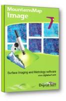
DIGITAL SURF
France
Dedicated image analysis software for optical microscopes and laboratory images MountainsMap® Image software makes it possible to: Add dimensions to your images and turn them into metrology data. Extract horizontal contours for geometric dimensioning. Quantify binary, grayscale and color-shaded spots, grains and particles. Enhance image quality and remove defects. Convert color images into 3D for better understanding of color variation. Link images with other types of surface data handled by Mountains®.
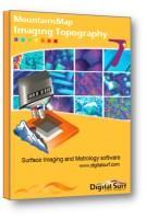
DIGITAL SURF
France
Dedicated surface imaging, analysis and metrology software for 3D optical microscopes and profilers measuring topography - confocal microscopes, interferometric microscopes, digital holographic microscopes, focus variation microscopes & structured light systems Compatible with all 3D optical profilers and microscopes used for surface analysis and metrology. Real time imaging of 3D surface topography with near-perfect lighting. 3D surface overlays for fast feature location - overlay color and intensity images on 3D topography. Remove data acquisition artifacts - outliers, local defects. Increase field of view virtually - assemble measurements using surface topography stitching. Analyze roughness and surface texture - in accordance with the latest ISO and national standards. Analyze surface geometry - including volume of surface structures (bumps, holes), step heights. Extract and analyze regions of interest - study them in the same way as full measured surfaces. 3D reconstruction of multi-focus images - reconstruct 3D topography from multi-focus image stacks. Easy integration into lab and production environments - export of all numerical results. Easy publication - export analysis documents, pages and individual images up to 1200 dpi. Add optional modules for advanced surface texture analysis, contour analysis, grains and particles analysis, 3D Fourier analysis, image co-localization, statistics and more.
NT-MDT LLC
Russia
Rebirth of Force Spectroscopy Advanced Nanomechanical, Electrical, Optical, Thermal and Piezoresponse Studies Fast Quantitative Nanomechanical Measurements and Force Volume Simultaneous Electrostatic and Nondestructive Conductivity, Piezoresponse and Thermal Studies Advanced CantileverType TipEnhanced Raman Scattering and Scanning NearField Optical Microscopy Topography in Attraction and Repulsive Regimes Young’s Modulus and Force Volume Adhesion and Work of Adhesion Conductivity InPlane and OutofPlane Piezoresponse Temperature and Thermal Conductivity Thermoelectric Electrostatic Kelvin Probe Force, Electrostatic Force and Scanning Capacitance Force Microscopy NearField Component of Optical Response TipEnhanced Raman Scattering In HybriD mode the tipsample distance is modulated according to the quasiharmonic law.
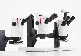
LEICA MICROSYSTEMS
Germany
Continuously improving production, keeping defect rates low, and fulfilling customer requests in order to stay competitive can be very challenging. Leica has developed the S9 stereo microscope series to help you cope with these challenges. With this new generation of Greenough stereo microscopes operators will be able to reveal details faster as they spend less time having to adjust the microscope. Available in different versions for diverse needs the S9 stereo microscopes boost efficiency and optimize optical inspections in your production line or Quality Control division, due to: - FusionOptics technology with 12 mm depth of field to find details fast - High magnification up to 55x and 9:1 zoom for quick changes from overview to details - 122 mm working distance for easy sample manipulations under the microscope - Integrated, network-camera for easy image sharing
Results for
Optical microscopes - Import exportNumber of results
16 ProductsCountries
Company type